Pics of Yeast under my new microscope

Homebrew Talk - Beer, Wine, Mead, & Cider Brewing Discussion Forum

Help Support Homebrew Talk - Beer, Wine, Mead, & Cider Brewing Discussion Forum:

This site may earn a commission from merchant affiliate links, including eBay, Amazon, and others.
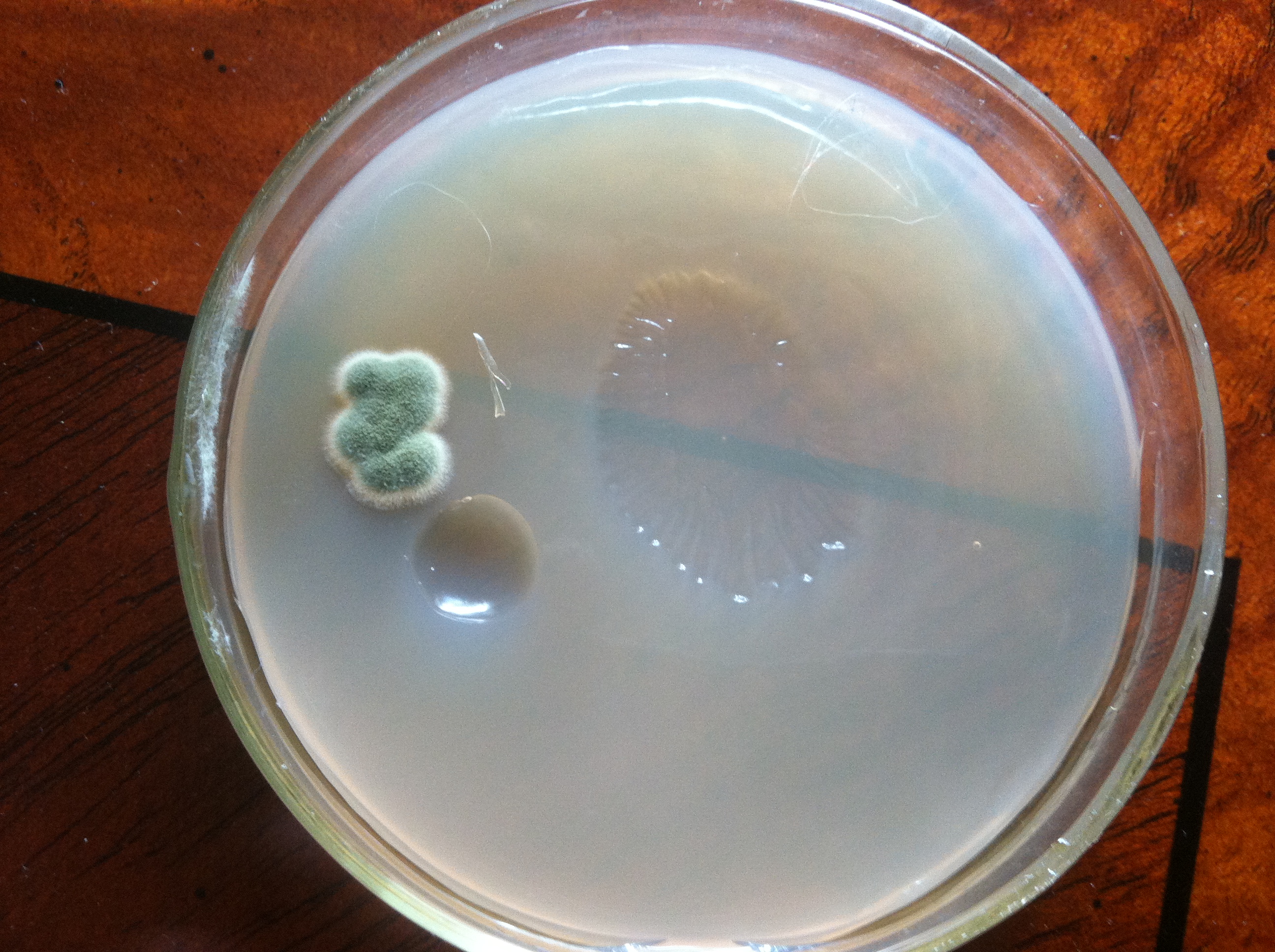
Picture 2 is a harvest from Lakefront Brewery's Wisconsinite from earlier this summer. Looks very different from 3068 and is much cleaner. Viability was determined from a different sample and was 6%. I might try stepping this culture up to see how it goes.

I used this culture in a 1 gallon batch and it went all sour/acetic on me. Nothing from the starter or the fermented batch looked bacterial, so maybe I got some wild yeast in there or the Jeremy King strain went wild.

I'm tuned in, let see some bacteria dance :ban:
 
I used this culture in a 1 gallon batch and it went all sour/acetic on me. Nothing from the starter or the fermented batch looked bacterial, so maybe I got some wild yeast in there or the Jeremy King strain went wild.

I'm tuned in, let see some bacteria dance :ban:

I'm jealous of those pics you put up there. I see very crisp images in my eyepieces, but I get some crap from this camera mounted on there. Not only is the resolution terrible, but I've got to keep playing with the RGB and exposure to get the pics to look right. I've got an IR filter mounted on the light source to help since the huge IR from the lamp causes all sorts of problems. Argh.

Glad someone is looking at this. I thought I was talking to myself again.
 
Surprised I've never noticed this thread! Very cool. I also enjoyed watching your Pool as Wort Chiller vid where you stood there wondering what to do with the overflowing carboy as your pooch licked off the wort chiller :D are you still doing this?
 
Surprised I've never noticed this thread! Very cool. I also enjoyed watching your Pool as Wort Chiller vid where you stood there wondering what to do with the overflowing carboy as your pooch licked off the wort chiller :D are you still doing this?

Bomber! No, my old IC is long retired (attic) much to Marlee's dismay.
 
passedpawn said:
Bomber! No, my old IC is long retired (attic) much to Marlee's dismay.

Marlee. I thought you called him Barley. Wouldn't be the worst name, just funny ;)
 
passedpawn, don't be jealous, it is a work microscope. You'd be wasting your money if you wanted it. I think your setup is pretty cool and all you need actually. It would be neat to have my own home yeast lab but I only get to brew 12 batches a year and fear it wouldn't be worth the extra trouble for me alone.
 
I found some decent time lapse software (VideoVelocity) that I just tested and works with camera I have mounted in my scope. So I should be able to do some better video of that sort. I want to record a sequence of yeast budding.

FYI, I'm probably going to stop updating this thread soon. It's reading mostly like a personal blog, which I already have for yeast and beer stuff. I know there's a few people interested, but it's mostly a one-way conversation here and not really appropriate for a forum. I'll post a link to my blog when I get it better organized.
 
Ok, this is sorta cool. I recorded 22 minutes of that bacteria growing across the slide. I then dropped that video into Pinnacle Studio and compressed it into 2 seconds, then uploaded to you tube.

http://www.youtube.com/watch?v=QTChFSQdcpo

Very interesting, I can picture this creating the streaked film on top of the beer. It's too bad it takes so long of video to show the growth. Great effort PassedPawn.
 
Very interesting, I can picture this creating the streaked film on top of the beer. It's too bad it takes so long of video to show the growth. Great effort PassedPawn.

This new software I'll be using this weekend will make the time lapse stuff much easier (no more video editing). It takes single snapshots at intervals, exactly what I need; i.e., one snapshot per minute over a day. Unfortunately it's going to cost me $35 (I tried some free utilities but they were not very impressive).

One problem I have is that during growth I have to occasionally adjust focus. I trust the X/Y stage of my scope, so I guess there is vertical movement of the media (agar substrate) somehow. The living culture might just be rising too, not sure yet. I had to adjust the focus a couple of times during that video and it created some weirdness in the final time lapse video.
 
How did the colonies of the lacto look from the berliner weiss yeast on the plate? Are they easily distinguishable with the naked eye vs. the sacc? I can't really see two distinct colony shapes on the picture of the plate you posted, but I figured they should look considerably different.
 
How did the colonies of the lacto look from the berliner weiss yeast on the plate? Are they easily distinguishable with the naked eye vs. the sacc? I can't really see two distinct colony shapes on the picture of the plate you posted, but I figured they should look considerably different.

The first pic in #198 was wild yeast I got from COLObrewer. I moved the pic out of that post.

So far, the colonies look the same, but the lacto colonies aren't pure. When I say it's lacto, what I meant is that it's slurry from a berliner weisse I made. That beer had both lacto and sacc in it. The plastic jar with the slurry has been sitting at room temp and now has a sweet pellicle on it. I plated from that pellicle. That plate is shown in the pics under the scope. I also thought the colonies looked like the other colonies I'm growing now, namely S-05 and COLObrewer mystery yeast.

So, while the plated lacto looks very similar to the sacc plates, it sure doesn't look like sacc under the scope. Honestly, I don't know what I'm doing here. I need to do more experiments to understand what to expect.

I plan on taking some pictures of these colonies under my lower power inspection scope and start to catelog this mess of information I'm accumulating.
 
It looks like when you're streaking your plates, you still have too much yeast on the innoculating loop (or whatever you are using) even at the end of the streaking, and you're not getting colonies forming from single cells. So the colonies are going to be mixed sacc and lacto and basically be a large colony all along the streak path. It looks like you're already streaking from where you've streaked cells into unstreaked agar each pass, so the streaks should get progressively fewer cells on each pass. Are you sterilizing your loop between passes? You should be able to get single colonies at least toward the end, where you'll have some colonies forming from single sacc cells and others forming from single lacto cells (or small clumps of several cells if they have really attached to each other). You should actually be able to isolate the sacc from the lacto on a properly streaked plate, or isolate sacc from a contaminated sample so you can use the purified sacc strain.

If you're actually plating from a pellicle, I'd guess that there wouldn't be much sacc in the pellicle, so you'd get mostly lacto colonies, but if you wanted to isolate and make sure you had some pure lacto, you could isolate from those individual colonies.
 
Thanks!

Yes, I don't have many (any ) single cell colonies. You're right, I'm putting too much down. I knew to be careful of that and I still did it.

I have a nichrome inoc loop and an alcohol lamp. I'm incinerating the loop, cooling on the agar, then dipping into my liquid cultures. Obviously I'm getting too much there.

I'm going to keep working on it. I'll get better. Again, thanks a lot for your advice.

It looks like when you're streaking your plates, you still have too much yeast on the innoculating loop (or whatever you are using) even at the end of the streaking, and you're not getting colonies forming from single cells. So the colonies are going to be mixed sacc and lacto and basically be a large colony all along the streak path. It looks like you're already streaking from where you've streaked cells into unstreaked agar each pass, so the streaks should get progressively fewer cells on each pass. Are you sterilizing your loop between passes? You should be able to get single colonies at least toward the end, where you'll have some colonies forming from single sacc cells and others forming from single lacto cells (or small clumps of several cells if they have really attached to each other). You should actually be able to isolate the sacc from the lacto on a properly streaked plate, or isolate sacc from a contaminated sample so you can use the purified sacc strain.

If you're actually plating from a pellicle, I'd guess that there wouldn't be much sacc in the pellicle, so you'd get mostly lacto colonies, but if you wanted to isolate and make sure you had some pure lacto, you could isolate from those individual colonies.
 
I'm lobbying that you don't move this to blog. It garners way more attention (expertise) being on the forum than it likely would on a blog. That benefits everyone. There's no shame in keeping journals on here, I follow several threads like that here such as the 100% Homegrown, and it's way more convenient to follow here than blog. My 2¢
 
I have a nichrome inoc loop and an alcohol lamp. I'm incinerating the loop, cooling on the agar, then dipping into my liquid cultures. Obviously I'm getting too much there.

So, after streaking an area of the plate with the loop, do you put the loop back in the flame to sterilize it, touch the agar, and then (without getting more liquid from your culture) streak another area of the plate? If not, you could try that. I see your streaks are overlapping a little for every time you change directions, so, it should pick up some yeast from the first streak and thin it out over the next streak, etc. I have only streaked a couple of plates, and they were by no means excellent, but if you aren't sterilizing the loop between streaks, give that a try to see if you get single colonies on one of the areas.

I also vote for you not to move this to a blog. I know I enjoyed seeing all your images so far, and I probably wouldn't have even seen them if they were in a blog instead.

I don't sterilize between. I just streak, then start with the tail of that streak and streak again. After 3 of those, I do one more in the middle. That should be the money streak. I just need more practice. The stuff grows really quickly so I'll know soon when I've mastered it. In fact, I think I'll streak a few plates right now.
 
I don't sterilize between. I just streak, then start with the tail of that streak and streak again. After 3 of those, I do one more in the middle. That should be the money streak. I just need more practice. The stuff grows really quickly so I'll know soon when I've mastered it. In fact, I think I'll streak a few plates right now.

Ah, that's the problem. You really need to sterilize or use a new loop each time. Here's a crude MSPaint drawing of what I mean. You're basically getting a tiny bit of your previous streak onto your loop each time, and spreading it out over a new section of your agar. By the time you get 3-4 passes in, you'll be guaranteed single colonies even if you start with a VERY dense liquid culture.

streaking.png

Alright. I'll try flaming the loop between streaks. That makes sense too.
 
If you aren't careful, you'll get airborne fungus / mold in your plates or slants. All of the unused slants I made are free from contamination. But one of the plates I made contracted a disease.

This plate had some odd craters in it, I guess from a bubble that was in there. I opened it up and looked at the craters under my inspection scope when it was a day old; I only looked at it for about 20 seconds. In that time, I guess something dropped out of the air. Here the plate is, about 10 days old.

full
 
Last edited:
I got a new toy for taking pictures in my scope, so I intend on adding more content here soon. I've made a bunch of plates, but I didn't need to make slants since I have a bunch of them that look perfect. Except one. The lid wasn't tight.

Anyone want to try to pick out the one that dried up :) For those familiar with marine fauna, dried agar agar media looks just like a nudibranch.

_mg_7953-62240.jpg
 
I picked up a cheap AmScope M152A and the 40x objective hits the hemocytometer when it focuses. This isn't normal, right? Anyone else have this issue?

It's not an immersion lens is it? the gap from lens to coverslip ought to be barely perceptible with those. It could be the haemocytometer is a little thick for the lens working distance.
 
Wow. What a read! Just got into propagating yeast and doing the whole cell counting thing here, and boy what a difference it makes! Yes, doing a starter really helped, but actually getting into the science... Thanks to all who contributed to this thread in the past... Even though it appears dead, I hope to add something of my own one day.

Seriously, this was the first thread outside the Boneyard where I read every single post. No shame in admitting I learned infinitely more here!

Prost!
 
There's a guy on ebay who refurbs Olympus microscopes.... he does an AMAZING job... Mikroscopes is his handle. I've bought 4 used scopes in the last year and returned all but his. There are tons of charlatans out there. Great thing is with ebay you can always send them back and seller pays shipping if they are not right.

Guy (Mikroscopes) used to work for Olympus back in the day. I contacted him via ebay and he built me something spec'd out exactly how I wanted then told. me when the listing was going up. Get a CHA or CHS model. This was the flagship lab line in the 80ies for them. 10x, 40x, 100x....Aplan objectives (edit - if you want phase contrast, objectives will be PL and have a position for the phase condenser/annulus like PH2, PH4, etc). For only cell counting you don't need it but when trying to identify stuff via morphology, it's really useful.

Amscope are garbage optics. If you're cell counting at 400x ONLY, they are fine.... but what will eventually happen is you'll have fun with the scope and want to do more.... that's when you need decent optics...

Just my 2cents.... oh and my Olympus cost 250$! 100, 400 and 1000x phase contrast and brightfield for 250 bucks with nice glass in a functionally new (yet 25 year old) scope.
 
Found this thread after some research on the subject, just about to order my hemocytometer and possibly some methylene violet if this vintage Sears 1200X microscope I found in my closet pans out. I'm skeptical about what my results will be just have to wait for the stain and polyurethane to dry (I made my new casing for the LED out of wood haha).

Old light was broken so quick trip to RadioShack and I now have a 9V system running a 10mm 3.5V 20mA LED which I'm assuming is bright enough.

But I loved reading every single post on this thread so helpful.

View attachment 1467264609027.jpg
 
Found this thread after some research on the subject, just about to order my hemocytometer and possibly some methylene violet if this vintage Sears 1200X microscope I found in my closet pans out. I'm skeptical about what my results will be just have to wait for the stain and polyurethane to dry (I made my new casing for the LED out of wood haha).

Old light was broken so quick trip to RadioShack and I now have a 9V system running a 10mm 3.5V 20mA LED which I'm assuming is bright enough.

But I loved reading every single post on this thread so helpful.

That LED you have there is a point source with a lens on it that does exactly the opposite of what you want - it disperses the light instead of condensing it. I don't think that LED is going to work very well. It might at very low magnification, but at higher levels you really need condenser optics below the stage.

The condenser matches the numerical aperture (NA) of the objective, thus getting the most possible light through the optics. See the pic below. "Abbe" condensers are the most common, and are on just about every consumer microscope sold. They allow you to manually control the NA (cone of light) to manually match the NA.

2000px-Condenser_diagram.svg.png
 

Latest posts

Back
Top